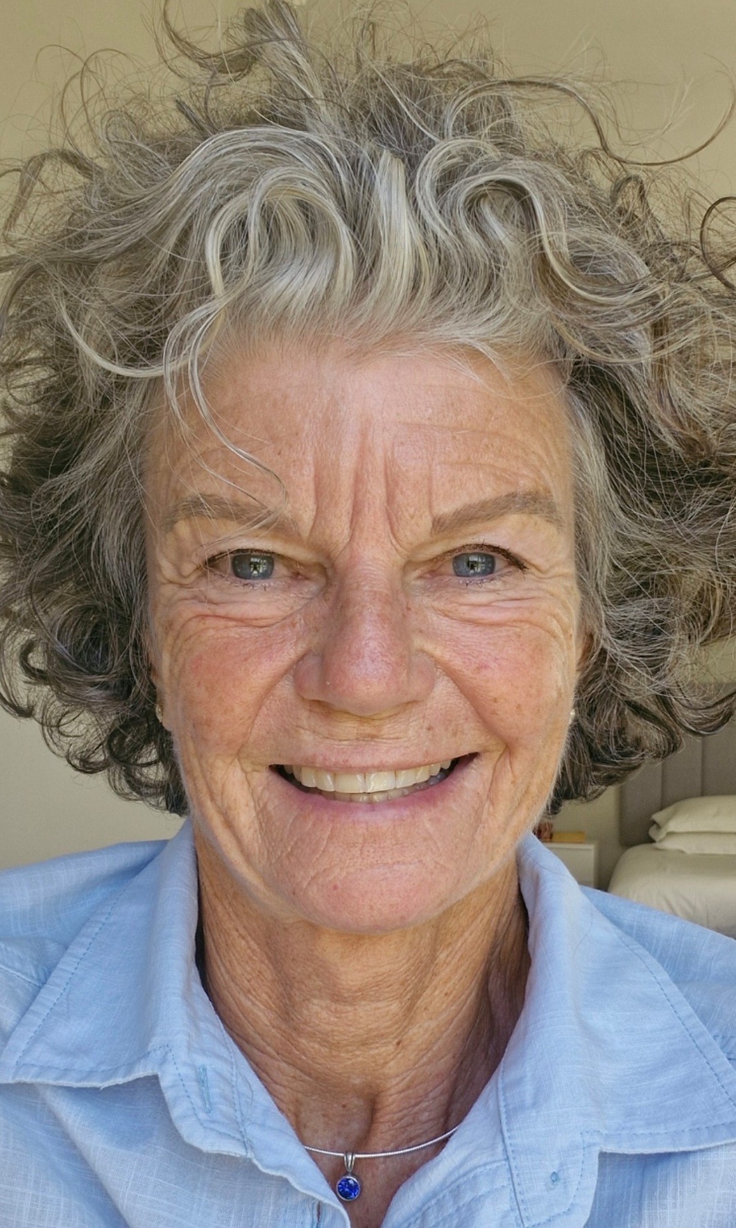
Linda shares her breast cancer journey

Linda was 45 years old when she was diagnosed with breast cancer. Like many women, there was no breast cancer in her family that she knew of, and she was a healthy person with a healthy lifestyle.
The small lump, which was diagnosed as Stage 1, was found by Linda after a dinner with friends, where a conversation about breast self-examinations took place. Never having done one before, Linda tried it out and found the small lump. This was in December 2008.
A GP visit was booked, followed by an attempted biopsy at a hospital. ‘Not the best experience,’ is how Linda put it – ‘being poked and prodded and with no biopsy concluded.’ An appointment with Dr Apffelstaedt was then recommended by her GP, and a biopsy taken at that first appointment.
On Friday, 19 December, Dr Apffelstaedt called to tell Linda that her results were positive, that she would need surgery, but could spend Christmas, New Year and her birthday with family and friends first.
Surgery was scheduled for 30 January 2009, where the sentinel node would be checked first before the lumps (more smaller lumps were also found in the right breast, which were found to be benign) were to be removed. With the node pronounced clear, a bilateral lumpectomy and reduction were performed.
Linda’s treatment journey started from there!
Diagnosed as low-risk, but with atypical cells found in the lumps, Linda had genetic testing to ascertain whether she should be treated as high-risk and include chemotherapy in her regimen. Based on the genetic testing results, chemotherapy was deemed to be unnecessary, and in April 2009, Linda started with 27 days of daily radiation, then oral Neophedan.
‘My physical challenges during treatment included hot flashes and night sweats, exacerbating my already bad sleeping pattern and creating mood swings. My estrogen level was sky high (my cancer was estrogen positive, so low levels are required), so Neophedan was stopped. Zoladex injections started, which lasted for 5 years. That ended 2014.’
Linda is very open about the emotional side of processing a diagnosis and going through treatment. ‘By the end of April, I felt emotionally drained. I was trying so hard to be positive for myself and Martin, my husband, and two boys, Josh, 15 and Sams, 12. I felt exhausted, I wasn’t sleeping and felt down. The continuous lack of sleep made me irritable, accompanied by mood swings. I wanted to be happy and feel thankful, but I really couldn’t on some days. It seemed out of my control. Finding the right meds for my situation took a bit of time, which I think made me more impatient with myself and my progress.’
Each woman’s breast cancer journey is unique. The tools that each person finds and uses are also unique. Linda believes that it’s important to be able to talk about your journey with someone who understands. ‘Talk to others who have travelled the cancer road. One small piece of advice or positivity can change everything. Ask questions until you are satisfied that you have the right answer to feel reassured.
The best therapy was my daily journal, which has kept all these answers true, as well as reminded me of whence I came. A long journey. The good days and the bad days. Keeping myself surrounded by friends to talk to and keeping busy doing things.’
Linda is cancer-free in 2025. When asked if there are any continuing psychological challenges, her response is: ‘Not anymore – now in 2025. I do have a moment or two when I go for my annual mammogram. There’s always that “what if” in the back of one’s mind, but so far all has been well.’
Linda’s advice
It’s a long journey that only you can make, and it’s lonely sometimes because one wants to be positive, to feel grateful and resume one’s former happy, healthy life. But the reality is, it’s you, your life, and this journey never ends. It will always be with you, it’s how you choose to live with it that is important.
Information on biopsies
Is a biopsy always necessary to make an accurate diagnosis?
To confirm the diagnosis of cancer in the breast, removal of a specimen from the lesion is required. This was previously often done by an open biopsy in theatre, but these days should be accomplished by a needle core biopsy under local anaesthesia.
What are the different types of biopsies?
Fine needle aspiration (FNA) uses a very thin needle to collect fluid or cells directly from the mass. Usually, the practitioner can perform this procedure while feeling the lump to help guide the needle. If a mass is seen on a mammogram, but the lump cannot be felt easily, in a specialised breast health centre. Ultrasound or computer-guided mammographic imaging is used to locate the mass and guide the position of the needle. The use of computer-guided mammography to locate the mass and help guide the position of the needle is called stereotactic needle biopsy.
If this procedure locates fluid, it is an indication that the lump is a cyst. If the procedure locates a solid mass, individual cells from the mass will be removed and sent to a laboratory for further analysis under the microscope.
Using FNA, mammography, and a clinical breast exam, a practitioner can determine with about 98% accuracy whether a lump is benign or malignant. If, however, there is still doubt, a core needle biopsy may be ordered. No anaesthesia is required for fine needle aspiration. A disadvantage of FNA is that so little tissue is removed that detailed analysis and typing of a cancer is not possible. Therefore, fine needle aspiration is used mainly for cystic masses.
Core needle biopsy is nowadays the preferred method to obtain a representative specimen from a solid mass. It removes a 3mm by 2cm core of tissue from the lesion. Ultrasound or stereotactic guidance may be used. Due to the size of the needle, local anaesthesia is required.
Next to the diagnosis of suspicious soft tissue masses, core needle biopsy is used for assessment of microcalcifications (microcalcifications are tiny calcium deposits that show up as fine white specks on a mammogram). If an experienced team finds micro-calcifications to be suspicious, a final diagnosis of cancer will be made in 3 to 4 out of 10 cases.
Incisional biopsy involves surgical removal of just a portion of the mass, which is sent to a laboratory for further analysis under the microscope.
Excisional biopsy involves surgical removal of the entire mass, which is sent to a laboratory for further analysis under the microscope. Both incisional and excisional biopsies are invasive, expensive and should only be necessary in exceptional cases to come to a diagnosis. In experienced breast centres, in more than 98 to 99% of cases, a core biopsy will deliver an accurate diagnosis.


